LA Magazine Los Angeles Magazine’s Best Physical Therapy Practice in LA

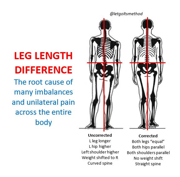
Leg Length Difference/Discrepancy

Ever had hips adjusted only for them to go out of alignment again? If so, then you likely have a structural leg length discrepancy (SLLD). While this may not seem like a big deal as SLLD is USUALLY negligible up to a point and doesn’t cause short-term problems by itself, it has a bottom-up effect on the body that can worsen over time, especially when coupled with other factors (e.g., stiffness, external trauma, stress from physical activities). When someone’s problems are chronic and unilateral (one-sided), they can often be traced back to a leg length discrepancy.

Due to the oblique angle of the pelvis, when one leg is longer than the other, it pushes the pelvis on that side up and back, which causes the opposite hip to “drop” and rotate forwards—in order to stand facing straight, the torso will rotate in the opposite direction.

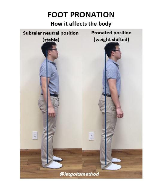
Since most SLLD’s involve a longer left leg, for explanation purposes, we’ll assume the left is always the longer side. When the left hip is higher than the right, the body tends to weight shift to the right—notice how the midline is much closer to the right leg in the image. In order to keep the center of gravity over the base of support, the lower back will curve to the left (this is a bit subtle in the image).  The upper back will then side-bend to the right, raising the left shoulder higher than the right. Finally, in order to keep the eye-line horizontal and remain upright, the head will tilt back to the left.

In general, this altered curvature of the spine causes the IT band, QL, obliques, scalenes, and levator scapulae to become tighter on the left than the right. On the right the hip flexors, piriformis, adductors, pectoralis and serratus anterior become tighter than the left, while the obliques, middle trap, and serratus posterior become weaker (reverse everything for someone with a longer right leg). Occasionally, this compensation pattern can also be altered by prior injuries or structural scoliosis.

Final point:

The only way to truly determine whether someone has SLLD is to measure the leg itself, from the greater trochanter of the femur to the fibular malleolus, and correlate it with the expected compensation pattern.

Pronation Part VII – How to Address Pronation II – Casting for Orthotics

As mentioned in our last post, many pairs of orthotics are made incorrectly and don’t address the main issue with pronation. Properly made orthotics must have support for the ball of the foot in order to properly address one’s pronation and the issues caused by it. If you haven’t already, go check out our previous pronation posts to get the relevant background information for this post!

Most issues with incorrectly-made orthotics can be traced back to the casting method—i.e., the method used to create the molds of the feet (which are then used to make orthotics). Many methods involve someone stepping into impression molds or onto pressure plates. The problem with this is that the individual is in a weight bearing position, which completely misses the reason behind pronation.

We cannot stress enough that pronation occurs because the ball of the foot must come down to the ground when weight bearing. Therefore, methods requiring someone to be in a weight bearing position will not result in accurate molds of the feet, as they won’t be in a subtalar neutral position. Proper casting methods require people to be in non-weight bearing positions in order to make orthotics properly. That said, many methods involving sitting positions also tend not to result in accurate foot molds: they often rely on active/passive dorsiflexion of the foot to hold the “neutral position” without accounting for the side-to-side rotation that occurs with pronation.

In our clinic, casting is done in a prone (lying face down) position. That way, the feet aren’t loaded and can be properly set (and held) in subtalar neutral while the casts are made. As seen in the video below, strips of plaster are used to encase the feet, which are held in subtalar neutral as the plaster sets. Based off of these molds, the orthotics will then be made with the proper amount of support to keep the ankle in subtalar neutral.

Pronation Part VII – How to address pronation – casting

One last, but very important, thing to remember about orthotics: since pronation is important for shock absorption, orthotics should still allow for some pronation to help with shock absorption, but not so much that the foot is constantly thrown out of neutral alignment.

Pronation Part VI – How to Address Pronation I – Orthotics

When people pronate, they’re often told to strengthen the foot muscles to “build an arch” that sets the foot in a neutral position. However, this assumes that pronation is the same as flat feet, which it is NOT. If you haven’t already, check out our previous pronation posts to get up to speed!

Remember, the issue with pronation is not with the arch, but with forefoot varus when the ankle is in subtalar neutral and the ball of the foot coming down to the ground when weight bearing. Therefore, training the muscles to create an arch helps minimally, as someone with forefoot varus will still pronate when the ball of the foot comes down. Even if the foot muscles can build an arch approximating subtalar neutral and are trained, they cannot maintain it 24-7: muscles eventually fatigue, causing the foot to pronate and the arch to collapse again—when dynamic movement is incorporated, that arch is even more likely to be lost.

So what corrects for pronation? The answer is orthotics: custom insoles made to keep the foot in a subtalar neutral position. This is achieved by supporting both the arch and the ball of the foot, such that it no longer drops when weight bearing—something that cannot be compensated for with foot musculature OR an arch support alone (pronation is a structural issue).

Many orthoses are actually just arch supports, but these don’t help much and may actually make the problem worse! Without support underneath the ball of the foot to maintain the forefoot varus position, pronation still occurs when weight bearing: the foot rolls over the arch support, which can further exacerbate one’s problems. Imagine shoving a rock underneath your foot and walking on it all day!

As seen in the photo, a pronated foot [1] still pronates on an orthotic that’s just arch support [2] because the ball of the foot is unsupported. However, with the properly made orthotics [3], the ball of the foot is supported so the foot no longer pronates and can maintain a subtalar neutral position. That said, there are some (rare) cases of calcaneal or midfoot instability (WITHOUT forefoot varus) that do benefit from arch supports alone. It all depends on the foot structure!

Pronation Part V – How Pronation Affects Low Extremity Movement II – Knee Flexion

Pronation 5_how pronation affects lower extremity movement_knee flexion

When the foot pronates and knee is rotated internally, any movement that requires flexion (bending) will occur at an oblique angle, as seen in the video above (the pronated foot is shown on the left, while the corrected foot is shown on the right). In other words, the knee does not move directly over the ankle, but rather off to the medial side (towards the midline of the body). Due to this oblique angle, people who pronate tend to excessively torque the knee—the degree of which depends on the degree of pronation. This puts additional strain on the anterior (front) and medial aspects of the knee, which may not be a significant issue when it comes to everyday activities such as walking, but it plays a huge factor in exercise and athletic performance.

Due to the additional torque on the knee caused by pronation, athletes in sports that involve explosive, rotational, and lateral movements—on top of rapid changes in direction—may be more prone to injuries. Since some individuals (particularly the young and/or fit crowd) may be able to compensate for their pronation, not necessarily everyone who pronates will experience symptoms of pain. However, regardless of injuries and/or pain, pronation can certainly affect athletic performance.

Having a “corrected” foot placed as close to subtalar neutral allows the knee and ankle to track properly, which translates to the optimal movement mechanics, as no energy is wasted in generating power. This significantly affects athletic performance, even in absence of injuries. When the knee doesn’t tract properly, more force is required to generate the same power and control the movement. This further exacerbates any issues caused by one’s pronation and can contribute to hip and knee stability. When the ankle and knee are properly aligned, the body is able to properly stabilize these structures and move more efficiently.

Pronation Part IV – How Pronation Affects Lower Extremity Movement I – Gait

In our first post regarding pronation, we discussed how pronation affects static posture. Today we’ll discuss how pronation affects dynamic movement, particularly the lower extremities. As the foot pronates and the midfoot rotates medially (inward), the knee also rotates internally. This alone may not seem like a big deal when dealing strictly with posture, but the real damage is occurs during movement.

When people pronate they tend to stand with their feet pointed outwards due to their internally rotated knees. In order to stand in a way that takes stress off the knees, the feet must point outward: if the feet are parallel, the knees will be at an oblique angle towards the midline, which is uncomfortable for most people. Therefore, to keep the knees pointing straight forward, the toes must turn out.

Pronation 4_how pronation affects lower extremity movement_gait

As a result, people who pronate tend to roll around the ball of the foot instead of rolling through it when they walk. As seen in the video, the pronated foot begins to roll inward (during the mid-stance phase of the gait cycle) before the heel leaves the ground, whereas the corrected foot remains stable. As the foot remains stable at the ankle and midfoot, it can then move through the ball of the foot instead of rolling around it (during the heel-off phase of the gait cycle). Due to this change in gait a person can develop any combination of the following issues: bunions, plantar fasciitis, neuromas, Achilles tendonitis, lateral ankle pain, knee pain, IT band or piriformis syndrome, low back or neck pain and poor posture and balance.

Pronation Part I – What is pronation I

Up till now, our posts have addressed stiff upper backs, tight muscles, and certain postures that can contribute to chronic neck and/or low back pain. However, what we haven’t addressed is WHY the thoracic spine gets stiff and WHY those muscles get tight. More specifically, what causes them to end up that way and what causes your body to end up in the same position time and time again.

The answer? YOUR FEET! More specifically, pronation: the inward rotation of the foot when weight bearing. This forces the weight onto the inside of the foot, as opposed to being evenly spread when the foot is in a subtalar neutral (stable) position, as seen in below:

Pronating also often comes with a flattening of the arches, which shouldn’t be confused with flat feet (to be covered later on). As the foot pronates, the knee rotates internally, (represented in the video links below).

Pronation 1_3_pronation anterior view

Pronation 1_4_pronation posterior view


When this happens on both sides of the body, the hips will drop into anterior pelvic tilt. Once that happens, the body’s weight will begin to shift forward. In order to keep ourselves from falling forward, the lower back will extend backwards to maintain the center of gravity. In order to keep the center of gravity in line with the hips, the thoracic spine becomes curved while the shoulders round out. More often than not, the head will also be shifted slightly forward (“turtleneck” position).

Here you can see how the body’s posture is affected by pronated feet vs feet set in subtalar neutral. The posture caused by pronation worsens over time as joints become stiffer and muscles become tighter. This chronic posture is why many people find it hard to simply sit or stand up “straight” and maintain that posture, as well as why many lower extremity injuries persist. It has nothing to do with how strong or weak the muscles are, but rather, with how their body compensates as a result of its structure – i.e., the bone structure of the foot having a bottom-up effect on areas higher up on the body.